Start-End dates: 01/09/20 – 31/08/23
Project reference: Retos Colaboración 2019 (RTC2019-006809-1)
Total budget: 940,000€
Financed by: State Research Agency under the Ministry of Science and Innovation, Spain

¨Funded by the State Research Agency under the Ministry of Science, Innovation and Universities; program Retos Colaboracion 2019, project reference: RTC2019-006809-1¨
Partners: Vall d’Hebron Research Institute (VHIR) (Barcelona, Spain)
Summary:
ZeClinics and Vall d’Hebron Institut de Recerca (VHIR) are proud to share significant progress in our mission to revolutionize cancer research through the utilization of Nanobodies.
Together, we have worked tirelessly to amplify the production of Nanobodies and assess their potential as powerful anti-oncogenic agents. The seamless coordination between ZeClinics and VHIR has been pivotal in managing and supervising this groundbreaking project.
Driven by a visionary goal, our efforts have been focused on demonstrating the remarkable capability of meticulously chosen nanobodies. These nanobodies have proven instrumental in impeding the relentless growth of both Cancer Stem Cells (CSC) and Differentiated Cancer Cells within our innovative zebrafish xenotransplantation model. From an extensive bacterial library, three extraordinary nanobody candidates have emerged, exhibiting potent anticancer properties.
Through the collaboration’s unwavering commitment, we’ve not only augmented production but also conducted thorough purification, subjecting these nanobodies to rigorous High-Performance Liquid Chromatography analysis to precisely identify their target interactions.
The outcome has been nothing short of extraordinary: a robust affinity for binding with CSC in vitro, coupled with a notable absence of adverse effects post-injection into the duct of Cuvier. This marks a significant milestone in our pursuit of a potential breakthrough in cancer treatment.
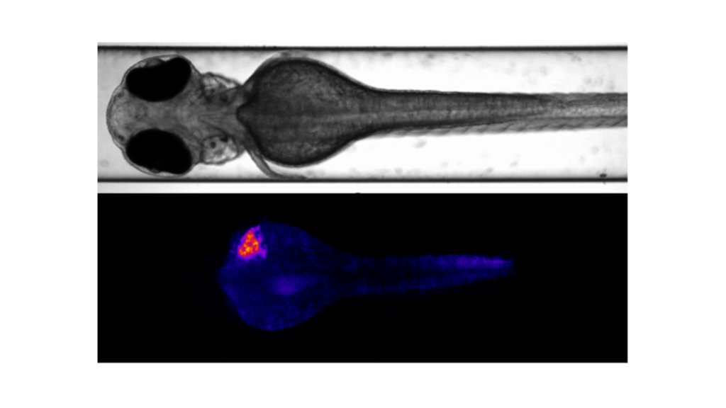 The larva of 48 hours post-fertilization (hpf) was injected into the perivitelline space with MDAMB-231 GFP-positive cells. The upper image displays a dorsal view captured with the Leica DM6 B in brightfield, presented in black and white. The lower image shows a dorsal view captured with the Leica DM6 B in the GFP channel, presented with a Look-Up Table (LUT).